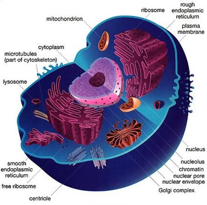

It’s also responsible for maintaining the cell’s shape.


Its function is to serve as a reservoir and maintain turgor pressure in the cell. Plant cells contain a large central vacuole that is full of water. But one of the major advantages was the ability of Nanolive Imaging to capture several biological phenomena and objects at the same time, enabling researchers to observe organelle rotation and other complex cellular dynamics involving multiple subcellular structures. Animal cells do not have a rigid cell wall, which is one of the reasons there are more cell types, organs, and tissues. The label-free, non-invasive imaging technique meant the team could observe the cells over an extended period of time. Taken together, the data paints a detailed picture of a cell’s structure and chemical composition, allowing scientists to precisely calculate dry mass, morphology, cell membrane dynamics and other features. They then quantified the observed phenomena using software specially designed to sift through the mass of raw imaging data. Purchase and download Amira software from ThermoScientific. Just like cells have membranes to hold everything in, these mini-organs are also bound in a double layer of phospholipids to insulate their little compartments within the larger cells. In this paper published in PLOS Biology in December 2019 by using Nanolive’s 3D Cell Explorer-fluo, researchers from EPFL could observe changes in cell size during division, organelle movements, and the formation of tiny lipid droplets – all over an extended period and without damaging the cell. High-resolution 3D images of organelles are of paramount importance in cellular biology. An organelle (think of it as a cell’s internal organ) is a membrane bound structure found within a cell.


 0 kommentar(er)
